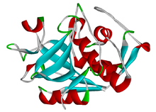
组织蛋白酶
| 组织蛋白酶 | |||||||||
|---|---|---|---|---|---|---|---|---|---|
 组织蛋白酶K的结构 | |||||||||
| 鑑定 | |||||||||
| 標誌 | CTP | ||||||||
| Pfam | PF00112(旧版) | ||||||||
| Pfam宗系 | CL0125(旧版) | ||||||||
| InterPro | IPR000668 | ||||||||
| SMART | Pept_C1 | ||||||||
| PROSITE | PDOC00126 | ||||||||
| MEROPS | C1 | ||||||||
| SCOP | 1aec / SUPFAM | ||||||||
| |||||||||
组织蛋白酶(英语:Cathepsin,缩写CTS)是存在于所有动物以及其他生物体中的蛋白酶(降解蛋白质的酶)。这个家族大约有十几个成员,它们的特点是它们的结构、催化机制以及切割的蛋白质。大多数成员可以在溶酶体中发现并在低pH值下被激活。因此,这个家族的活动几乎完全在细胞器中。然而也有例外,例如组织蛋白酶K,它在破骨细胞分泌后在骨吸收过程中在细胞外起作用。组织蛋白酶在哺乳动物细胞更新中起着至关重要的作用。
分类
[编辑]临床意义
[编辑]组织蛋白酶参与了许多生理过程,与许多人类疾病有关。半胱氨酸蛋白酶作为药物靶点已吸引了大量研究工作。[1][2]
- 癌症,组织蛋白酶D是一种丝裂原,它会减弱衰变趋化因子的抗肿瘤免疫反应,从而抑制树突状细胞的功能。[3]
- 中风[4]
- 创伤性脑损伤[5]
- 阿尔茨海默病[6]
- 关节炎[7]
- 埃博拉,已发现组织蛋白酶B和较小程度的组织蛋白酶L是病毒进入宿主细胞所必需的。[8]
- 慢性阻塞性肺病
- 慢性牙周炎
- 胰腺炎
- 几种眼部疾病:圆锥角膜,视网膜脱离,黄斑变性和青光眼[9]
组织蛋白酶A
[编辑]这种蛋白质的缺乏与多种形式的神经氨酸酶中毒有关。恶性黑色素瘤转移病灶裂解物中的组织蛋白酶A活性显着高于主要焦点裂解物中的组织蛋白酶A活性。组织蛋白酶A在被肌肉萎缩症和去神经疾病影响的肌肉中增加。
组织蛋白酶B
[编辑]组织蛋白酶B可作为β分泌酶1发挥作用,切割前类淀粉蛋白质以产生β淀粉样蛋白。[10]作为肽酶C1家族成员的编码蛋白的过表达,它与食管癌和其他肿瘤有关。[11]组织蛋白酶B还与包括卵巢癌在内的各种人类肿瘤的进展有关。
组织蛋白酶D
[编辑]组织蛋白酶D(一种天冬氨酰蛋白酶)似乎可以切割多种底物,如纤连蛋白和层粘连蛋白。与其他一些组织蛋白酶不同,组织蛋白酶D在中性pH值下具有一些蛋白酶活性。[12]肿瘤细胞中这种酶的高水平似乎与更大的侵袭性有关。
组织蛋白酶K
[编辑]组织蛋白酶K是最有效的哺乳动物胶原酶。组织蛋白酶K与骨质疏松症有关,骨质疏松症是一种骨密度降低导致骨折风险增加的疾病。破骨细胞是身体的骨吸收细胞,他们分泌组织蛋白酶K来分解胶原蛋白,它是骨头的非矿物质蛋白基质的主要成分。[13]除其他组织蛋白酶外,组织蛋白酶K通过细胞外基质的降解在癌症转移中发挥作用。[14]在患有动脉粥样硬化的老鼠中,组织蛋白酶S和K的基因敲除被证明可以减少动脉粥样硬化病变的大小。[15]组织蛋白酶K在培养的内皮细胞中的展现受剪切应力的调节。[16]组织蛋白酶K也被证明在关节炎中起作用。[17]
组织蛋白酶V
[编辑]老鼠的组织蛋白酶L与人类的组织蛋白酶V同源。[18]老鼠的组织蛋白酶L已被证明在老鼠的脂肪生成和葡萄糖耐受不良中发挥作用。组织蛋白酶L降解纤连蛋白、胰岛素受体(IR)和胰岛素样生长因子1受体(IGF-1R)。组织蛋白酶L缺陷的老鼠的脂肪组织较少,越低的血清葡萄糖和胰岛素水平,越高的胰岛素受体亚单位,葡萄糖转运蛋白(GLUT4)和比野生型更多的纤连蛋白。[19]
抑制剂
[编辑]五种环肽对人的组织蛋白酶L、B、H和K具有抑制活性。[20]几种抑制剂已经进入临床试验,以组织蛋白酶K和S为目标,有望成为治疗骨质疏松症、骨关节炎和慢性疼痛的药物。由于不良副作用,组织蛋白酶K抑制剂Relacatib、Balicatib和Odanacatib分别在I、II和III期的临床试验期间终止。[21]SAR114137是一种组织蛋白酶S抑制剂,在慢性疼痛的I期之后没有进展。2022年,组织蛋白酶L抑制剂STI-1558获得美国食品药品监督管理局(FDA)批准,开始治疗2019冠状病毒病(COVID-19)的I期研究。[22]
组织蛋白酶酶谱法
[编辑]酶谱法是一种凝胶电泳,它使用与底物聚合的聚丙烯酰胺凝胶来检测酶活性。组织蛋白酶酶谱法基于通过与明胶底物聚合的聚丙烯酰胺凝胶的迁移来分离不同的组织蛋白酶。电泳在非还原条件下进行,使用亮肽素保护酶免于变性。[23]确定蛋白质浓度后,将等量的组织蛋白质加入到凝胶中。然后让蛋白质迁移通过凝胶。电泳后,将凝胶放入复性缓冲液中,以使组织蛋白酶恢复其天然构象。然后将凝胶放入特定pH值的活化缓冲液中,并在37°C下孵育并放置一夜。该活化步骤允许组织蛋白酶降解明胶底物。当凝胶使用考马斯亮蓝染色剂染色时,仍含有明胶的凝胶区域呈现蓝色。组织蛋白酶活跃的凝胶区域显示为白色条带。该组织蛋白酶酶谱法已用于检测飞摩尔量的成熟组织蛋白酶K。可以根据它们的迁移距离基于它们的分子量来识别不同的组织蛋白酶:组织蛋白酶K(~37kDa)、V(~35kDa)、S(~25kDa)和 L(~20kDa)。组织蛋白酶具有特定的pH水平,在该水平下它们具有最佳蛋白水解活性。组织蛋白酶K能够在pH值为7和8下降解明胶,但这些pH值不能让组织蛋白酶L和V有很高的活性。相反,在pH值为4时,组织蛋白酶V具有活性,但组织蛋白酶K没有。调整活化缓冲液的pH值可以进一步识别组织蛋白酶类型。[24]
名称来源
[编辑]组织蛋白酶名称来源于古希腊语,由kata-(倒下)和hepsein(煮沸)合并称为Cathepsin。
参考文献
[编辑]- ^ Turk, Vito; Stoka, Veronika; Vasiljeva, Olga; Renko, Miha; Sun, Tao; Turk, Boris; Turk, Dušan. Cysteine cathepsins: From structure, function and regulation to new frontiers. Biochimica et Biophysica Acta (BBA) - Proteins and Proteomics. Proteolysis 50 years after the discovery of lysosome. 2012-01-01, 1824 (1) [2022-09-10]. ISSN 1570-9639. doi:10.1016/j.bbapap.2011.10.002. (原始内容存档于2022-10-27) (英语).
- ^ Turk, Vito; Stoka, Veronika; Vasiljeva, Olga; Renko, Miha; Sun, Tao; Turk, Boris; Turk, Dušan. Cysteine cathepsins: From structure, function and regulation to new frontiers. Biochimica et Biophysica Acta (BBA) - Proteins and Proteomics. January 2012, 1824 (1): 68–88. PMC 7105208
 . PMID 22024571. doi:10.1016/j.bbapap.2011.10.002.
- ^ Nomura T, Katunuma N. Involvement of cathepsins in the invasion, metastasis and proliferation of cancer cells (PDF). J. Med. Invest. February 2005, 52 (1–2): 1–9 [2022-12-10]. PMID 15751268. doi:10.2152/jmi.52.1
 . (原始内容存档 (PDF)于2021-04-27).
- ^ Lipton, Peter. Ischemic Cell Death in Brain Neurons. Physiological Reviews. 1999-01-10, 79 (4) [2022-09-10]. ISSN 0031-9333. doi:10.1152/physrev.1999.79.4.1431. (原始内容存档于2022-09-11).
- ^ Xu, Jianguo; Wang, Handong; Ding, Ke; Lu, Xinyu; Li, Tao; Wang, Jiawei; Wang, Chunxi; Wang, Jian. Inhibition of Cathepsin S Produces Neuroprotective Effects after Traumatic Brain Injury in Mice. Mediators of Inflammation. 2013, 2013 [2022-09-10]. ISSN 0962-9351. PMC 3824312
 . PMID 24282339. doi:10.1155/2013/187873. (原始内容存档于2022-09-10).
- ^ Yamashima, Tetsumori. Reconsider Alzheimer's disease by the 'calpain–cathepsin hypothesis'—A perspective review. 2013-06-01 [2022-09-10]. doi:10.1016/j.pneurobio.2013.02.004. (原始内容存档于2022-09-22).
- ^ Raptis, Sofia Z.; Shapiro, Steven D.; Simmons, Pamela M.; Cheng, Alec M.; Pham, Christine T. N. Serine Protease Cathepsin G Regulates Adhesion-Dependent Neutrophil Effector Functions by Modulating Integrin Clustering. Immunity. 2005-06-01, 22 (6) [2022-09-10]. ISSN 1074-7613. PMID 15963783. doi:10.1016/j.immuni.2005.03.015. (原始内容存档于2014-03-23) (英语).
- ^ Chandran, Kartik; Sullivan, Nancy J.; Felbor, Ute; Whelan, Sean P.; Cunningham, James M. Endosomal Proteolysis of the Ebola Virus Glycoprotein Is Necessary for Infection. Science (New York, N.Y.). 2005-06-10, 308 (5728) [2022-09-10]. ISSN 0036-8075. PMC 4797943
 . PMID 15831716. doi:10.1126/science.1110656. (原始内容存档于2022-09-10).
- ^ Im, Eunok; Kazlauskas, Andrius. The role of cathepsins in ocular physiology and pathology. Experimental Eye Research. 2007-03-01, 84 (3). ISSN 0014-4835. doi:10.1016/j.exer.2006.05.017 (英语).
- ^ Hook, Gregory; Hook, Vivian; Kindy, Mark. The cysteine protease inhibitor, E64d, reduces brain amyloid-β and improves memory deficits in Alzheimer’s disease animal models by inhibiting cathepsin B, but not BACE1, β-secretase activity. Journal of Alzheimer's disease : JAD. 2011, 26 (2) [2022-09-10]. ISSN 1387-2877. PMC 4317342
 . PMID 21613740. doi:10.3233/JAD-2011-110101. (原始内容存档于2022-11-30).
- ^ Habibollahi, Peiman; Figueiredo, Jose-Luiz; Heidari, Pedram; Dulak, Austin M; Imamura, Yu; Bass, Adam J.; Ogino, Shuji; Chan, Andrew T; Mahmood, Umar. Optical Imaging with a Cathepsin B Activated Probe for the Enhanced Detection of Esophageal Adenocarcinoma by Dual Channel Fluorescent Upper GI Endoscopy. Theranostics. 2012-02-16, 2 (2) [2022-09-10]. ISSN 1838-7640. PMC 3296470
 . PMID 22400064. doi:10.7150/thno.4088. (原始内容存档于2022-09-10).
- ^ 存档副本. journals.biologists.com. [2022-09-10]. (原始内容存档于2022-09-22).
- ^ Shi GP, Chapman HA, Bhairi SM, DeLeeuw C, Reddy VY, Weiss SJ. Molecular cloning of human cathepsin O, a novel endoproteinase and homologue of rabbit OC2 (PDF). FEBS Lett. January 1995, 357 (2): 129–34. PMID 7805878. S2CID 28099876. doi:10.1016/0014-5793(94)01349-6
 . hdl:2027.42/116965.
- ^ Gocheva, Vasilena; Joyce, Johanna A. Cysteine Cathepsins and the Cutting Edge of Cancer Invasion. Cell Cycle. 2007-01-01, 6 (1). ISSN 1538-4101. PMID 17245112. doi:10.4161/cc.6.1.3669.
- ^ Lutgens, E.; Lutgens, S.p.m.; Faber, B.c.g.; Heeneman, S.; Gijbels, M.m.j.; de Winther, M.p.j.; Frederik, P.; van der Made, I.; Daugherty, A.; Sijbers, A.m.; Fisher, A. Disruption of the Cathepsin K Gene Reduces Atherosclerosis Progression and Induces Plaque Fibrosis but Accelerates Macrophage Foam Cell Formation. Circulation. 2006-01-03, 113 (1) [2022-09-10]. doi:10.1161/CIRCULATIONAHA.105.561449. (原始内容存档于2022-08-02).
- ^ Platt, Manu O.; Ankeny, Randall F.; Shi, Guo-Ping; Weiss, Daiana; Vega, J. D.; Taylor, W. R.; Jo, Hanjoong. Expression of cathepsin K is regulated by shear stress in cultured endothelial cells and is increased in endothelium in human atherosclerosis. American Journal of Physiology-Heart and Circulatory Physiology. 2007-03-01, 292 (3) [2022-09-10]. ISSN 0363-6135. doi:10.1152/ajpheart.00954.2006. (原始内容存档于2022-09-11).
- ^ Salminen-Mankonen, H. J.; Morko, J.; Vuorio, E. Role of Cathepsin K in Normal Joints and in the Development of Arthritis. Current Drug Targets. [2022-09-10]. (原始内容存档于2022-09-10) (英语).
- ^ Brömme, Dieter; Li, Zhenqiang; Barnes, Michel; Mehler, Ernest. Human Cathepsin V Functional Expression, Tissue Distribution, Electrostatic Surface Potential, Enzymatic Characterization, and Chromosomal Localization. Biochemistry. 1999-02-01, 38 (8) [2022-09-10]. ISSN 0006-2960. doi:10.1021/bi982175f. (原始内容存档于2022-10-31) (英语).
- ^ Weinstock, David M.; Brunet, Erika; Jasin, Maria. Formation of NHEJ-derived reciprocal chromosomal translocations does not require Ku70. Nature cell biology. 2007-08, 9 (8) [2022-09-10]. ISSN 1465-7392. PMC 3065497
 . PMID 17643113. doi:10.1038/ncb1624. (原始内容存档于2022-09-10).
- ^ Bratkovič, Tomaž; Lunder, Mojca; Popovič, Tatjana; Kreft, Samo; Turk, Boris; Štrukelj, Borut; Urleb, Uroš. Affinity selection to papain yields potent peptide inhibitors of cathepsins L, B, H, and K. Biochemical and Biophysical Research Communications. 2005-07-08, 332 (3). ISSN 0006-291X. doi:10.1016/j.bbrc.2005.05.028 (英语).
- ^ Mullard, Asher. Merck & Co. drops osteoporosis drug odanacatib. Nature Reviews Drug Discovery. 2016-10-01, 15 (10) [2022-09-10]. ISSN 1474-1784. doi:10.1038/nrd.2016.207. (原始内容存档于2022-09-01) (英语).
- ^ Cooley, Brian. "Sorrento Therapeutics Announces the FDA IND Clearance of STI-1558, An Oral M(pro) and Cathepsin L Inhibitor to Treat COVID-19". Sorrento Therapeutics, Inc. July 19, 2022 [September 1, 2022]. (原始内容存档于2022-12-10).
- ^ Li, Weiwei A.; Barry, Zachary T.; Cohen, Joshua D.; Wilder, Catera L.; Deeds, Rebecca J.; Keegan, Philip M.; Platt, Manu O. Detection of femtomole quantities of mature cathepsin K with zymography. Analytical Biochemistry. 2010-06-01, 401 (1). ISSN 0003-2697. doi:10.1016/j.ab.2010.02.035 (英语).
- ^ Wilder, Catera L.; Park, Keon-Young; Keegan, Philip M.; Platt, Manu O. Manipulating substrate and pH in zymogr aphy protocols selectively distinguishes cathepsins K, L, S, and V activity in cells and tissues. Archives of biochemistry and biophysics. 2011-12-01, 516 (1) [2022-09-10]. ISSN 0003-9861. PMC 3221864
 . PMID 21982919. doi:10.1016/j.abb.2011.09.009. (原始内容存档于2022-09-10).
外部链接
[编辑]- The MEROPS online database for peptidases and their inhibitors: A01.010[失效連結]
- 醫學主題詞表(MeSH):Cathepsins
